Necrosis
It is defined as a localized area of dead tissue followed by degradation of tissue by hydrolytic enzymes liberated from dead cells, it is invariably accomplished by an inflammatory reaction. Necrosis: Pathology, Types & Causes- Healthpathy

Etiology:
Necrosis caused by various agents-
- Hypoxia:- most common and it is due to the reduced supply of blood to the cell.
- Physical agents:- due to mechanical thermal trauma, electricity, and radiations.
- Chemical agents:- chemical poisons such as Hg, Ar, Cyanide and strong acids, salt of glucose, and
oxygen at high concentrations. - Microbial Agent:- Infection caused by virus, bacteria, fungus, etc.
- Immundiblelogical Injury:- Due to hypersensitive reaction and anaphylactic reaction.
Pathogenesis:
In a type of necrosis irreversible cell injury is characterized by two essential changes as follows-
A. Cell Digestion by Lytic Enzymes:-
Morphologically change is identified as a homogeneous and intensely eosinophilic cytoplasm. It may show cytoplasmic vacuolation or cytoplasmic calcification.
B. Denaturation of Protein:-
Morphologically characterized by nuclear changes by necrotic cells. The nuclear changes include Condensation of nuclear chromatin, which may either undergo dislocation or fragmentation, into many clumps.
Necrosis: Pathology, Types & Causes- Healthpathy
Types of Necrosis:-

Morphologically it is characterized as follows –
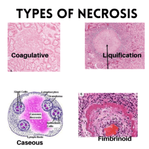
1. Coagulative Necrosis:-
- It is the most common type of Necrosis caused by irreversible focal injury mostly from the sudden association of blood flow and less often from bacterial and chemical energy.
- Commonly affected organ:-
a. Kidney
b. Heart and spleen. - Grossly:- In the early stage foci of coagulative necrosis is pale, firm, and slightly swollen and with progression, they become yellowish, soften, and shrunken.
- Microscopically:-
- The Hallmark of coagulative Necrosis is the condensation of normal cells into
their ‘Tomb Stone ‘ i.e outline of cells are retained so cell types can still be recognized but there
cytoplasmic and nuclear details are lost. - Necrosed cells were swollen and appears more eosinophilic along with nuclear changes.
- Eventually, the necrosed focus is inflicted by inflammatory cells, and the dead cells are
phagocytosed by leaving granular debris and fragments of cells.
2. Liquification Necrosis:-
- It occurs due to the degradation of the tissue by the action of powerful hydrolytic enzymes.
- Common Examples are:- Infract brain and Abscess cavity.
- Grossly:- The affected area is soft with a liquified center containing necrosis debris. Later a cyst wall is formed.
- Microscopically:-
- The cystic space contains necrosis calls debris and macrophages fitted with phagocytized material.
- The cyst wall is formed by perforating capillaries, inflammatory cells, and gliosis, in the case of brain Infract proliferating fibroblasts – in the case of an abscess cavity.
3. Caseous Necrosis:-
- It is found in the center of the foci of tuberculosis infections and has combined features of both coagulative and liquefactive necrosis.
- Grossly:- Foci of caseous necrosis resembles a dry cheese and are soft granular and yellowish.
- Microscopically:-
- The necrosed foci are structureless, eosinophilic, and contain granular debris.
- The surrounding tissue shows characteristic granular inflammatory reaction consisting of epithelial cells with expressed giant cells of laughs and foreign body type and peripheral mantle of lymphocytes.
4. Fat Necrosis:-
- It is a special form of cell death occurring at two anatomically different locations but Morphologically similar lesions. These are –
a. Acute pancreatic necrosis
b. Traumatic fat nacrosis. - Grossly:- Fat necrosis appears as yellowish-white and firm deposits.
- Microscopically:-
- The necrosed fat cells have a cloudy appearance and are surrounded by an inflammatory reaction.
- The formation of calcium soaps is identified in the tissue section as the amorphous granular and
basophilic material.
5. Fimbrinoid Necrosis:-
- It is characterized by the deposition of fimbrin-like material which has the staining properties of fimbrin.
- It is found in various Examples of immunologic tissue injury e.g.- vasculitis, autoimmune disease, Arthur reaction, etc.
- Microscopically:- It is identified by brightly eosinophilic hyaline-like deposition in the vessel
wall.
Frequently Asked Questions
Necrosis: Pathology, Types & Causes- Healthpathy
How to differentiate necrosis from apoptosis?
Apoptosis is a predefine programmed cell death for the smooth functioning of the body. It is also known as programmed cell death whereas necrosis is a localized area of dead tissue due to some external causes such as injury, radiation, or chemical exposure.
How to identify necrosis?
Necrosis can be identified as-
- Swelling of the cell
- Swelling of the nucleus
- Karyolysis (nuclear dissolution)
- Karyorrhexis (nuclear fragmentation)
- Nuclear pyknosis
- Pale eosinophilic cytoplasm
- Cytoplasmic vacuoles may be present in areas of necrosis
- Adjacent cellular debris and inflammatory cells may be present if there is a leakage of the cell membrane
What is TNF?
Tumor necrosis factor (TNF) is an adipokine and a cytokine. It is mainly produced by activated macrophages, T lymphocytes, and natural killer (NK) cells. It can induce the person’s immunity.
Is necrosis good or bad?Necrosis: Pathology, Types & Causes- Healthpathy
Necrosis indicates the death of body tissue which may vary in different conditions, but in some cases, it can have poor prognoses such as in the cases of tumor cancer.
Does necrosis reversible?
Necrosis indicates cell death which means permanent cell death that is irreversible while reversible cell injury mainly occurs mainly in cases of ischemia.
More to study-


Follow us on-


